
Each urodynamic test has a distinct diagnostic purpose. Additionally, there are multiple ways to perform each assessment, which can affect the test’s results. Here, the merits of ultrasound versus catheterization for measuring post-void residual1 (PVR) urine volume will be discussed.
Post-Void Residual Urine Assessment
 The PVR test examines how much urine is left in the bladder after voiding. The amount of residual urine (leftover) can easily be measured by using ultrasound or by draining the bladder with a thin flexible tube (catheter). The latter comes with a slight risk of infection or injury, but it is safe when performed carefully. And it may be less expensive than its counterpart: ultrasound.
The PVR test can be used to diagnose a number of disorders. These include:
- Incontinence
- Enlarged prostate
- Bladder stones
- Bladder obstruction
- Urethral stricture
- Constipation
- Infection
- Neurological disorder
- Weak bladder muscle
In general, a normal PVR volume for healthy patients younger than 65 years of age is less than 50 mL. For older patients, PVR greater than 100 mL is considered abnormal. This test is simple to perform, and involves measuring the volume of urine that remains in the bladder after the patient urinates. There are two methods for measuring PVR: ultrasound, and catheterization. Both options have advantages and disadvantages.
Ultrasound
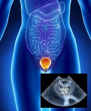
After the patient voids his or her bladder, an ultrasound4 can be performed at the patient’s bedside. This non-invasive procedure simply requires a bladder scanner, which is a portable 2D or 3D ultrasound bladder scanning device. A portable scanner is comprised of a base unit with a display screen and an ultrasound transducer. The scanner emits ultrasound waves and the returning echo appears as a cross section of the bladder. Bladder and PVR measurements are automatically calculated. While catheterization was once the norm for PVR measurement, ultrasound is now considered to be the preferred option thanks to the numerous advantages of its use.
- Advantages
The advantages of ultrasound are diverse. For patients, there is less discomfort surrounding the procedure, as an ultrasound is non-invasive and causes no pain or handling of genitals. There is no risk of complication, such as urinary tract infection or damage, and an ultrasound is less time consuming. In comparison to catheterization, ultrasound is quick and easy to perform, which requires less time for hospital staff to complete the examination.
- Disadvantages
However, it is important to correctly interpret the results of a bladder scan. Accuracy2 is the key, and ultrasound is not perfect. In some instances, cystic lesions3 (read more here on urodynamics and interstitial cystitis) in the pelvis have been misidentified as post-void residual urine. Additionally, bladder scanners must be properly maintained and calibrated in order to provide the most accurate results. Since PVR is automatically determined by the bladder scanner, it is important that care is taken to fully understand the variables that affect these measurements. In addition, this procedure is typically more expensive than simple catheterization.
Straight Catheterization
Alternatively an in and out catheter5 can be used to drain any remaining urine in the bladder for measurement. Before bedside bladder scanners became more widely available, catheterization was the primary method for determining PVR. While bladder ultrasound is recommended in most cases, there are certain patient conditions in which a catheter is required. These include:
- Morbid obesity
- Severe abdominal scarring
- In situ abdominal staples or tension sutures
- Infected abdominal wound
- Pregnancy
- For children aged <36 months
- Early postpartum
- Advantages
The advantages of this technique primarily include cost-effectiveness and versatility for instances where a bladder scanner is not appropriate, or is unavailable. In addition, there is no concern of misinterpreting the results of a catheterization for a PVR assessment.
There is no additional maintenance required for keeping catheterization equipment in working order, as is the case for a bladder scanner and the results require no specialized interpretation. Perhaps most importantly, bladder scans can reveal when catheterization is required, so starting with catheterization can eliminate unnecessary procedures.
When PVR is calculated in context of a urodynamics test, catheterization is preferred because the catheter is already in place to support the urodynamics testing.
- Disadvantages
The disadvantages of PVR measured via catheterization are material. Catheterization is invasive and increases the risk of urinary tract infection. There is significantly more preparation time, due to the necessity of a sterile working environment. Patients experience some discomfort, as this procedure requires the insertion of a catheter. Finally, catheterization can only be performed in certain settings, and does not have the same portability as a bladder scanner.
Final Thoughts
Ultimately, use of a bladder scanner4 is often preferred in order to reduce the risk of complications; however, extreme care must be taken to ensure accurate results with this method. Ultrasound is definitely the way to go if the patient has some anxiety about the use of a catheter, while the catheterization can be used to drain any remaining urine in the bladder for measurement. Both have their advantages and disadvantages, and it's up to the medical professional to choose the best option.

References
- Asimakopoulos, A. D., Nunzio, C. D., Kocjancic, E., Tubaro, A., Rosier, P. F., & Finazzi-Agrò, E. (2014). Measurement of post-void residual urine. Neurourology and Urodynamics, 35(1), 55-57. doi:10.1002/nau.22671 Link
- Haylen, B. T., & Lee, J. (2008). The accuracy of post-void residual measurement in women. International Urogynecology Journal, 19(5), 603-606. doi:10.1007/s00192-008-0568-0 Link
- Alagiakrishnan, K., & Valpreda, M. (2009). Ultrasound bladder scanner presents falsely elevated postvoid residual volumes. Canadian Family Physician, 55(2), 163–164. Link
- Teng, C., Huang, Y., Kuo, B., & Bih, L. (2005). Application of Portable Ultrasound Scanners in the Measurement of Post-Void Residual Urine. Journal of Nursing Research, 13(3), 216-224. doi:10.1097/01.jnr.0000387543.68383.a0 Link
- Jalbani, I. K., & Ather, M. H. (2014). The accuracy of three-dimensional bladder ultrasonography in determining the residual urinary volume compared with conventional catheterisation. Arab Journal of Urology, 12(3), 209–213. http://doi.org/10.1016/j.aju.2014.05.001 Link