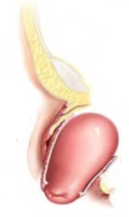
 Pelvic Organ Prolapse (POP) is a condition for which it is estimated that up to 11% of women1 will undergo surgery in order to correct associated symptoms, such as urinary incontinence, by the time they reach 85 years of age. Many tools are used for diagnosing POP, one of which is urodynamic assessment. Here, Pelvic Organ Prolapse and urodynamics will be extensively discussed.
What is Pelvic Organ Prolapse?
Pelvic Organ Prolapse2 is a condition characterized by a prolapse of one or more pelvic organs. When pelvic organs, such as the bladder, urethra, uterus, vagina, small bowel, or rectum move out of their customary positions, they can place pressure on other organs or body parts and cause discomfort, pain, urinary incontinence, and other undesirable side effects. One or more pelvic organs can be prolapsed at the same time. The many types of POP include:
Bladder
Bladder prolapse (also known as cystocele) is a condition that occurs when the surrounding tissue and muscles which hold the bladder in place become weakened or stretched. It can occur due to cancer or due to other degenerative diseases. When cystocele occurs, the bladder will move from its standard position and press against the vagina. In this case, a bulge can be felt through the vaginal wall3.
Urethra
In a urethral prolapse (urethrocele), the muscles and tissue that hold the urethra in place become weak. As a result, the urethra can both curve and widen. Women who experience a urethral prolapse commonly also experience a bladder prolapse at the same time. Like a bladder prolapse, a urethra prolapse can press against the vaginal wall. Latest research on this particular health issue is on young girls4.
Uterus
Uterine prolapse5 is caused by the weakening of the pelvic muscles and ligaments. In this instance, the uterus drops from its normal position and the cervix bulges down, into the vagina.
Vagina
A vaginal vault prolapse commonly occurs after the removal of the uterus. When the top of the vagina is no longer supported by the uterus, the unsupported portion can drop into the vaginal canal.
Small Bowel
A small bowel prolapse (also called enterocele) arises when the tissues and muscles that support this organ are weakened or stretched. The small bowel moves from its normal position and presses against the vaginal wall.
Rectum
A rectum prolapse5 (also called a rectocele) occurs when tissues and muscles that support the end of the large intestine are weakened or stretched. In this instance the rectum will move from its normal position and press against the back vaginal wall. If vaginal walls are weak, the rectum might also bulge into the vagina.
What are the Symptoms of Pelvic Organ Prolapse?
The most common symptoms of POP involve sensations of pain or weakness in the pelvic area. Women experiencing pelvic organ prolapse often feel pressure against the vaginal wall when the pelvic organs press or bulge against the vagina. Women may also experience a “full” feeling in the lower abdomen.
A patient might describe feeling as though something is falling out of her vagina, such as a tampon, or pain in the lower back or groin. Urinary incontinence and frequent urination are also common symptoms, as are constipation and fecal incontinence. Finally, vaginal pain during intercourse is another common complaint for someone experiencing POP.
What Causes Pelvic Organ Prolapse?
There are many common reasons a woman might experience POP. The two most common risk factors for Pelvic Organ Prolapse are childbirth and hysterectomy. During pregnancy and delivery, the muscles and tissues that support a woman’s pelvic organs are stretched. This phenomenon can lead to weakened muscles as well as organ displacement, resulting in prolapse. However, a cesarean section is not strongly linked with POP.
After a hysterectomy, the removal of the uterus can leave surrounding organs with less natural support, as in the case of a vaginal prolapse. Additional risk factors for POP include obesity, a chronic cough, frequent constipation, and pelvic organ tumors. Each of these factors place additional stress on the pelvic organs and can cause them unnaturally stretch or weaken.
Paralysis disorders that affect the pelvic floor muscles can also be linked with increased incidence of POP. Multiple sclerosis, muscular dystrophy, and spinal cord injury all commonly result in POP.
Research suggests that age and family history are also linked with POP. Women going through menopause have lower estrogen levels, which affects collagen production. Collagen is important for support in the pelvic connective tissues; therefore, less collagen can lead to pelvic organ displacement.
How is Pelvic Organ Prolapse Diagnosed?
Pelvic Organ Prolapse can be diagnosed during a routine medical exam where medical history, reported symptoms, pregnancy history, and pelvic exam results are considered. Additional tests that can be ordered to elucidate the findings include cystoscopy, intravenous pyleogram, CT scan, and urodynamics. A cystoscopy will determine whether a bladder or urethral prolapse has occurred. Intravenous pyelogram will show the size, shape, and position of pelvic organs. A CT scan can be used to provide a detailed picture of the pelvic area. Finally, urodynamic tests will provide a clear view as to how the body stores and releases urine.
After POP is diagnosed, a classification6 is given to describe the organ’s level of prolapse. While many classification systems exist, the most common method is to stage the prolapse based on closeness of the lowest part of the prolapsed organ to the opening of the vagina. A high staging level (i.e. III or IV) denotes a more advanced case.
How is Pelvic Organ Prolapse Treated?
Pelvic Organ Prolapse can be treated in a number of ways, ranging from non-surgical to surgical. A woman with mild POP symptoms can make lifestyle changes that include pelvic floor exercises or use of a removable pessary to support prolapsed organs.
Women experiencing pain, difficulties with bowel or bladder function, or interference with sexual activity should consider surgery. Surgical methods include repair of supporting tissues near the prolapsed organ or vaginal wall, or even hysterectomy.
Pelvic Organ Prolapse and Urodynamics
Now that POP symptoms, diagnosis, and treatment have been outlined above, the next section of this article will specifically discuss POP and the role of urodynamics in diagnosing the condition and identifying post-surgical complications.
What are Urodynamics?
Urodynamic tests are considered the gold standard in diagnosing problems with the lower urinary tract. A series of tests are performed to characterize the functioning of the bladder and urethra for elucidation of problems such as incontinence, bladder obstruction, and even cancer.
First, a urine flow test is typically performed to determine characteristics of a patient’s urine stream. Next, two catheters are inserted into the patient, one into the urethra and another into the vagina or rectum, and the bladder is filled and voided. This test looks for bladder obstruction, muscle weakness, stress induced incontinence, urethra strength, and other disorders that affect the pelvic organs, muscles, and tissues.
Other assessments include urethral pressure profilometer, which measures the strength of contractions in the sphincter; electromyography, which measures electrical activity in the bladder neck; and fluoroscopy, which is a video x-ray of the pelvic organs.
When are Urodynamic Tests Ordered for Pelvic Organ Prolapse?
The International Scientific Committee of the Third International Consultation on Urinary Incontinence has stated that urodynamic testing is highly recommended7 for any woman who is seeking surgical treatment for incontinence due to pelvic organ prolapse.
Urodynamic tests can determine whether an obstruction is being experienced that is caused by POP. Flow rate and pressure/flow studies are integral in this determination. Indeed, urodynamic level of outflow that is used for defining obstruction is more sensitive in women than in men, making this test particularly attractive for use in female patients suffering from Pelvic Organ Prolapse. Additionally, video urodynamics can also help in the staging of POP, particularly when bladder or urethral prolapse is suspected.
What Types of Urodynamic Abnormalities are observed with Pelvic Organ Prolapse?
Bladder obstruction is among the most common reasons to order urodynamic testing for POP. Bladder outlet obstruction can be defined as a low maximum free flow rate of less than 12 mL/s that persists for the patient in combination with high detrusor pressure greater than 20 cm H2O during a pressure-uroflow study. Indeed, approximately 24% of women8 who test positive for bladder outlet obstruction according to these standards experience severe forms of POP.
In one study, urodynamic testing was shown to be a good indicator of post-operative urinary conditions9 in POP patients. Clinical records of 87 POP patients who underwent surgery to repair their disorders were examined. Pre-operatively, each woman underwent a cough stress test and a pressure flow study. After POP reduction, these same women were evaluated using uroflowmetry, postvoid residual determination, and symptom assessment via a questionnaire.
Following surgery, detrusor over-activity was found to be a good predictor of post-operative urgency to urinate and urge urinary incontinence. Immediately following surgery postvoid residuals increased, but returned to a normal level within 1 month of surgery. The best predictor of large postvoid residual reoccurrence was poor detrusor contractility immediately following surgery.
Therefore, the researchers from this study recommend post-operative urodynamic evaluations including a cough stress test for stress urinary incontinence, as well as a test for detrusor function. These assessments will help predict post-operative urinary conditions for POP patients.
Is Urodynamic Assessment Necessary for Pelvic Organ Prolapse?
A recent study published in European Urology10 sought to answer the question of whether urodynamic assessment is necessary for diagnosing stress urinary incontinence in conjunction with POP, particularly relative to a powerful prediction model and artificial neural network (ANN).
In the study, ANN technology was used to elucidate the connection between urinary symptoms and urodynamic assessment in women experiencing POP. The goal of this research was to determine whether ANN could supplement or replace pre-operative urodynamic testing for women with POP. Of 802 women tested, significant associations were found between baseline data, symptoms, anatomic observations, and urodynamic diagnosis. Ultimately, researchers determined that, while ANN is powerful, urodynamic testing is the best way to evaluate pre-operative patients at this time, and that urodynamics should be “par for the course” during urinary dysfunction assessment.
In another study on this issue, published in Urologic Clinics of North America11, the utility of urodynamics for POP was examined. Ultimately, there were four main points from this study, both advocating for and against urodynamic assessment as mandatory for pre-operative POP patients.
The first point was that urodynamics is an important tool for identifying stress incontinence following surgery. One of the main side effects of prolapse reduction is stress incontinence, yet many women are reticent to talk to their doctor about symptoms. Including urodynamic testing as part of the post-POP reduction routine could help to immediately recognize this issue.
Next, it was determined that urodynamic testing can be beneficial for developing a plan to manage symptoms of patients who are clinically continent. This is especially true if both patient and physician are on board with selective management of the urethra during POP surgery.
The authors of this study warn that the usefulness of urodynamic assessment is limited in patients that experience both POP and symptoms of overactive bladder. Here, urodynamic testing will not be beneficial for elucidating pre- or post-operative problems with high selectivity.
Finally, the authors of this study assert that urodynamic testing should be performed on an individual basis for POP patients. The cost effectiveness of these studies – for both patient and clinic – is not immediately known. Additionally, there is no clearly defined answer at present as to how the results of urodynamic testing impact patient counseling or treatment. At this time, urodynamic assessment is still very much an art – as opposed to a science – when it comes to POP reduction.
What is the Usefulness of Urodynamic Assessment for Pelvic Organ Prolapse?
A recent study examined the urodynamic changes in POP12 and the role of urodynamic testing in helping surgeons prevent post-operative stress urinary incontinence. Here 50 cases of advanced (stage III) pelvic organ prolapse were studied.
Patients were given a pre-operative cystometrography (CMG) to assess filling and voiding phases of the bladder. The women then underwent surgical correction for POP via vaginal hysterectomy with pelvic floor repair. Following surgery, all women received a post-operative CMG within four weeks of surgery.
The most common symptoms that women with advanced POP reported were urinary frequency, urgency, and difficulty voiding. After surgery, fewer patients experienced these symptoms, except for diurnal frequency, which increased (possibly due to surgery). Data from the CMGs showed that pre-operatively, the first desire to void occurred at a higher bladder volume (235 mL) than post-operatively (203 mL). The normal desire to void also decreased, while bladder capacity increased pre- and post-operation.
The most significant changes were observed during the voiding phase. Max flow rate increased post-operatively (18.6 mL/s versus 9.2 mL/s). Detrusor pressure at peak flow decreased post-operatively as well (24.6 cm of water versus 34.4 cm of water). Residual volume post-void decreased significantly post-surgery (94.6 mL versus 149.3 mL), as did detrusor opening pressures (18 cm of water versus 29.7 cm of water).
The most important takeaway message from this data is that 68% of advanced POP patients show mild outlet obstruction during urodynamic assessment. Additionally, the higher post-operative urine flow rates show that surgical correction of POP relieves the symptoms of bladder obstruction. Finally, maximum flow rate during voiding phase is uniformly low for patients both pre- and post-operation.
The conclusions that the researchers put forth here is that urodynamic testing has indicated that the urodynamic profile of a woman experiencing advanced POP improves after surgery. Urodynamic evaluation is not recommended by these researchers unless the patient also experiences stress urinary incontinence. In this instance, a CMG is recommended in order to identify intrinsic sphincter deficiency, which can be repaired during POP surgery.
Summary
Ultimately, Pelvic Organ Prolapse and Urodynamics go hand in hand. Pelvic Organ Prolapse is estimated to affect up to 11% of women at some point in their lifetime. This disorder most commonly affects women who have given childbirth or have had a hysterectomy, but risk factors also include obesity, smoking, pelvic tumors, and neural disease. Urodynamic assessment for POP is still being studied. Researchers have pointed out both positive and negative evaluations for its use. Ultimately, urodynamic assessment should be performed prior to POP reduction surgery, particularly if stress urinary incontinence is also reported.
If you are looking for a way to provide urodynamics cost effectively in your practice, click the button below.
References
- Panicker, R., & Srinivas, S. (2009). Urodynamic Changes in Pelvic Organ Prolapse and the Role of Surgery. Medical Journal Armed Forces India, 65(3), 221-224. doi:10.1016/s0377-1237(09)80007-5 Link
- Schmid, C., & Maher, C. F. (2013). Epidemiology, Risk Factors, and Social Impact of Pelvic Organ Prolapse. Pelvic Organ Prolapse, 1-9. doi:10.1016/b978-1-4160-6266-0.00001-0 Link
- Smith, P. P., & Appell, R. A. (2006). Pelvic organ prolapse and the lower urinary tract: The relationship of vaginal prolapse to stress urinary incontinence. Current Bladder Dysfunction Reports, 1(1), 19-26. doi:10.1007/s11884-006-0003-7 Link
- Holbrook, C., & Misra, D. (2011). Surgical management of urethral prolapse in girls: 13 years' experience. BJU International, 110(1), 132-134. doi:10.1111/j.1464-410x.2011.10752.x Link
- Scherer, R., Marti, L., & Hetzer, F. H. (2008). Perineal Stapled Prolapse Resection: A New Procedure for External Rectal Prolapse. Diseases of the Colon & Rectum, 51(11), 1727-1730. doi:10.1007/s10350-008-9423-0 Link
- Persu, C., Chapple, C., Cauni, V., Gutue, S., & Geavlete, P. (2011). Pelvic Organ Prolapse Quantification System (POP–Q) – a new era in pelvic prolapse staging . Journal of Medicine and Life, 4(1), 75–81. Link
- Abrams P, Andersson KE, Birder L, Brubaker L, Cardozo L, Chapple C, Cottenden A, Davila W, de Ridder D, Dmochowski R, Drake M, Dubeau C, Fry C, Hanno P, Smith JH, Herschorn S, Hosker G, Kelleher C, Koelbl H, Khoury S, Madoff R, Milsom I, Moore K, Newman D, Nitti V, Norton C, Nygaard I, Payne C, Smith A, Staskin D, Tekgul S, Thuroff J, Tubaro A, Vodusek D, Wein A, Wyndaele JJ. Members of Committees; Fourth International Consultation on Incontinence. Fourth International Consultation on Incontinence Recommendations of the International Scientific Committee: Evaluation and treatment of urinary incontinence, pelvic organ prolapse, and fecal incontinence. Neurourol Urodyn. 2010;29:213–240. Link
- Groutz, A., Blaivas, J. G., & Chaikin, D. C. (2000). Bladder outlet obstruction in women: Definition and characteristics. Neurourology and Urodynamics, 19(3), 213-220. doi:10.1002/(sici)1520-6777(2000)19:3<213::aid-nau2>3.0.co;2-u Link
- Araki, I., Haneda, Y., Mikami, Y., & Takeda, M. (2009). Incontinence and detrusor dysfunction associated with pelvic organ prolapse: clinical value of preoperative urodynamic evaluation. International Urogynecology Journal, 20(11), 1301-1306. doi:10.1007/s00192-009-0954-2 Link
- Costantini, E., & Lazzeri, M. (2011). Urodynamics for Pelvic Organ Prolapse Surgery: “Par for the Course”. European Urology, 60(2), 261-262. doi:10.1016/j.eururo.2011.04.001 Link
- Ballert KN. Urodynamics in pelvic organ prolapse: when are they helpful and how do we use them? Urol Clin North Am. 2014;41(3):409–17. doi: 10.1016/j.ucl.2014.04.001. Link
- Panicker, R., & Srinivas, S. (2009). Urodynamic Changes in Pelvic Organ Prolapse and the Role of Surgery. Medical Journal Armed Forces India, 65(3), 221-224. doi:10.1016/s0377-1237(09)80007-5 Link


